Ventricular Tachycardia Ablation
What is Idiopathic Ventricular Tachycardia?
In some hearts, an abnormal heart rhythm develops in the bottom part of the heart when an electrical impulse starts from a different location. This impulse may cause the heart to have extra beats or start racing. Certain things in some people can trigger episodes. These include caffeine, alcohol, anxiety, exercise or sudden movements such as bending over. However, often these episodes can occur at any time without a trigger. During an episode, you may be aware of the beating of your heart. Other symptoms might include dizziness (blackout out may occur but is unusual), shortness of breath, and anxiety. Many people have no symptoms. Why this occurs is not known.

Is Idiopathic Ventricular Tachycardia dangerous?
In the vast majority of cases Idiopathic VT is a benign condition. This means that it will not cause sudden death or a heart attack. It will not shorten life expectancy. There are some rare exceptions that will be discussed with you if relevant. In some people frequent extra beats can affect the heart’s function.
What treatments are available for Idiopathic VT?
There are 3 main options for people with Idiopathic VT:
- No treatment at all. Because Idiopathic VT is a benign condition, for those people having infrequent and short-lived episodes that are not troublesome one option is to simply live with it.
- Medication. For people who do not wish to continue having episodes a second option is to take regular daily medication. There are a variety of different possible medications. Medications reduce the frequency and severity of episodes but do not cure the problem. There is also the possibility of developing side effects from these drugs.
- Radiofrequency Ablation. This is a procedure that cures the condition.
What is Radiofrequency Ablation (RFA)?
Radiofrequency is a low power, high frequency energy that causes a tiny region of the heart near the tip of the catheter to increase in temperature, thus ablating (or cauterising) a small area of abnormal tissue. Radiofrequency energy has been used for decades by surgeons to cut tissue or to stop bleeding. For the treatment of palpitations, a much lower power of radiofrequency energy is used.
What happens during the ablation procedure?
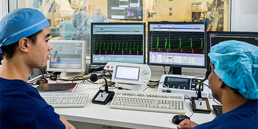
You will usually be admitted to hospital on the day of your procedure. You will be required to fast for at least six hours before the study. Prior to the procedure you will require an ECG. Once in the Electrophysiology Laboratory (EP Lab) you will be given a light sedative and your groin will be shaved. The EP lab has a patient table, X-Ray tube, ECG monitors and various equipment. The staff in the lab will all be dressed in hospital theatre clothes. Many ECG monitoring electrodes will be attached to your chest area and patches to your chest and back. These patches may momentarily feel cool on your skin.
A nurse or doctor will insert an intravenous line usually into the back of your hand. This is needed as a reliable way to give you medications during the study without further injections. You will also be given further sedation if and as required. You will also have a blood-pressure cuff attached to your arm that will automatically inflate at various times throughout the procedure. The oxygen level of your blood will also be measured during the EP study and a small plastic device will be fitted on your finger for this purpose. Your groin area and possible your neck will be washed with an antiseptic cleansing liquid and you will be covered with sterile sheets leaving these areas exposed.
The procedure may be performed under local anaesthetic with sedative medication or occasionally under full general anaesthetic. This will be discussed with you before the procedure. If the procedure is performed under local anaesthetic, the doctor will inject the anaesthetic to the area in the groin where the catheters are to be placed. After that, you may feel pressure as the doctor inserts the catheters but you should not feel pain. If there is any discomfort you should tell the nursing staff so that more local anaesthetic and sedative medication can be given. Occasionally it is also necessary to place a catheter in a vein in the side of the neck.
The catheters are positioned in your heart using X-Ray guidance. Once the catheters are in place you may feel your heart being stimulated and usually your abnormal heart rhythm will be induced. When the type of abnormal rhythm has been identified and the abnormal tissue localised, the radiofrequency ablation will be applied to this spot. This may cause a transient warm discomfort in the chest.
Radiofrequency ablation procedures are lengthy and the average duration is approximately 2 to 3 hours.
What are the Risks Of An Idiopathic VT Ablation Procedure?
Although most people undergoing Radiofrequency ablation do not experience any complications, you should be aware of the following risks.
- Local bleeding, blood clot or haematoma (blood collection) – this may occur at the catheter insertion site.
- Rapid abnormal heart rhythm – this may actually cause you to pass out for a very short period of time and in some cases a small electric shock may be required to restore your normal rhythm.
- Perforation or damage – very slight chance this may occur to either a heart chamber or to the wall of one of the arteries.
- Heart block – depending on the location and type of your abnormal rhythm being ablated, there is a chance of damage occurring to the heart’s normal electrical system (the AV node). This may be temporary, but permanent damage would result in a permanent pacemaker being inserted. This would have to be performed immediately at the time of the procedure.
- Major complications – worsening heart function, stroke, heart attack and death are rare.
- Other rare complications (reported in the literature) include: haemo or pneumothorax (blood or air in the chest wall requiring tube drainage) and damage to the phrenic nerve (the nerve supplying the diaphragm).
Radio-frequency ablation is an effective and safe way to cure patients suffering from Idiopathic Ventricular Tachycardia.
